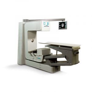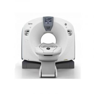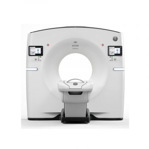


Description
Millions of patients around the world are still waiting to receive the standard of care they need and in an affordable way. Healthcare providers are seeking better technology to help more patients and drive better outcomes. Simple and fast, CT is arguably the most valuable diagnostic imaging tool in healthcare. Yet its capacity to improve patient care is far from tapped. All revolutions start somewhere. Our revolution began with the Revolution CT system — designed from the ground up for pioneering the future of CT. Now we introduce Revolution ACT. Revolution ACT is designed not only as a product, but as a solution to help you provide better patient care — and provide it to more patients. It’s the centerpiece of our commitment to help you achieve your CT services aims.
You’ll find many of the technologies inherited from our most advanced Revolution CT system – like the new Clarity panel detector, new user interface, or Organ Dose Modulation. Helping you feel more confident about moving to the next level, clinically and economically — that’s intelligent innovation.
Revolution ACT: It’s exactly what’s needed to help you achieve success. And exactly what you’d expect from GE.
Clarity Panel Detector
Highly innovative segmented panel designs built with advanced packaging and miniaturization technology for lower power consumption and improved thermal performance. Since thermal control is localized on the integrated panel, the detector achieves better precision in performance and reduces the detector startup time.
The higher density of interconnects and modern A/D chips deliver better signal linearity with fast response time. An integrated DAS on Detector or DoD design, the analog signal transmission length from Detector to DAS is cut to a virtual zero reducing noise by up to 20% and improving image quality
Overlapped Reconstruction
A higher sampling density provides better representation of the original signal and can potentially lead to improved image quality and fidelity. The term “conjugate samples” refers to two projection samples that are 180º apart in their respective projection angle, but at an equal distance to the isocenter. Taking both sets of projection measurements into consideration, we obtain a significantly increased sampling density and, therefore, potentially improve the overall acquisition. Using GE’s novel conjugate cone beam reconstruction algorithm, the conjugated rows of projections are considered jointly in the back projection step and delivers improved Z-axis visualization performance. Conjugate cone-beam acquisition enables 32-slice imaging on Revolution ACT.
Ultra Kernel
Adaptive Enhance Level Adjustment (AELA) can improve visual spatial resolution while maintaining pixel noise standard deviation and artifact. This kernel maybe helpful in enhancing the visualization of small anatomical structures with high contrast. Combine with sub millimeter slice thickness acquisition, Revolution ACT allows imaging at higher spatial resolution and greater detail of small
Smart Dose with ASiR
ASiR uses statistical modeling to remove noise in images while preserving anatomical detail. This enables a same- or better-quality image to be produced with lower tube current or tube voltage, thus lower dose, for all CT applications in which it is used. ASiR is one of the key dose reduction technologies flowing down from our premium product line. ASiR is the most-used dose-lowering software in the industry. It allows healthcare providers to lower dose by up to 40% to their patients as compared to standard image reconstruction without reducing imaging quality. ASiR also improves low-contrast detectability and can have equivalent IQ to an acquisition with 1.67 times the mA2. Operators can manage longer coverage scans easier. The maximum tube power and current of ASiR allows achieving the same image quality at a lower mA with less tube heat output, which enables the tube for longer duration under helical scan. Revolution ACT performance will be equivalent to 40KW and 333mA (@120kV, 3.3MHu tube heat capacity equivalence with ASiR). This improved efficiency helps lower power consumption and operational costs to the administrators.
Smart Dose: Organ Dose Modulation (ODM)
Organ Dose Modulation (ODM) is a scan mode that was developed to reduce dose via x-ray tube current modulation for superficial tissues such as breasts, eyes, gonads etc. ODM was developed to provide the dose reduction goal of a shield material without the negative effects it may have on imaging performance. ODM not only reduces the dose to the sensitive organ by up to 40% but also optimizes the overall dose while maintaining the noise index (NI) value selected by the user.
Smart Dose: Advanced Noise Reduction Tech. (ANRT)
ANRT Is a reconstruction technology used to reduce the noise of diagnostic images and retain detailed information of the image structure. ANRT noise-reduction technology can, while maintaining image noise level, lessen the dose of some X-ray testing, or improve image noise level and image quality. ANRT is only applicable on digital oblique images.
Smart Dose: Volumetric image Space Recon (ViSR)
Volumetric Image Space Reconstruction (ViSR) helps reduce artifacts and noise without compromising resolution. It contains an adaptive filter that is designed in the projection space with the filter parameters dynamically adjusted to adapt to the local noise characteristics such that the amount of smoothing operation is based on the x-ray flux level at each projection channel. In anatomical regions such as the brain, this 3D filter reduces noise without compromising resolution, for clear visualization of brain, tumor, and pediatric cases. With ViSR the scanner delivers up to 20% image quality improvement at the same dose, or the same image quality with up to 36% dose reduction.
Smart Flow: pitch booster with IQ Enhance
IQ Enhance is an advanced algorithm to reduce helical artifact in thin-slice helical scanning. The Revolution ACT scanner with this feature can accelerate its helical pitch up to 3.0x when acquiring the same helical artifact level compared with the same scanner with IQ Enhance disabled. Use of IQ enhance (IQE) along with sub-second rotation speed allows faster pitch scanning covering more anatomy at similar image quality.
Smart Flow: new user interface
Smart Flow with new user interface: the workflow and user interface have been completely redesigned with continuous feedback from a cross section of technologists and radiologists to make it easy-to-learn and use. The workflow features a modern look and feel that is inspired by our Revolution CT product. Many steps of the user experience have been reworked right from patient entry, to protocol selection, to monitoring and processing the patient scan. Smart new features include a simple and advanced mode – where users can switch between the modes at the click of a button.
Smart Flow: volume helical digital tilt
Volume helical digital tilt is an innovation in image reconstruction technology that allows clinicians to reconstruct tilted views of up to 30 degrees without the need for physically tilting the scanner. Digital tilt uses a novel resampling technology within the reconstruction pipeline to map vertical data samples to user specified tilted view angle. Digital tilt exams are more efficient since operators can setup the entire workflow from their console without the need for multiple trips between console and gantry controller. With a volume helical acquisition, clinicians have the additional advantage of leveraging powerful post processing and visualization tools for creating volume rendering, multiplanar reformatting (MPR), and curved MPR views as needed.
Smart Flow: spectral calibration
Spectral calibration calculates the beam hardening vectors based on a theoretical model of x-ray attenuation through beam-path and the data from scanning a 20 cm water phantom. Applying the result from spectral calibration in reconstruction, the uniformity of the CT numbers in image, especially the off-center image, is improved compared with the conventional beam hardening correction method.
Specifications
Gantry specifications
| Aperture | 65 cm |
| Maximum SFOV | 43 cm |
| Focal spot to detector distance | 94.9 + 0.1 cm |
| Tilt | + 30 degrees (digital) |
| Rotation Speed | 0.6, 0.98, 1.0, 1.2, 1.5, 2.0, 3.0, 4.0 seconds |
| Laser Alignment | Internal & external with + 1 mm accuracy |
Tube
| Anode Heat Storage | 2.0 MHU and 3.3MHU equivalent with ASiR |
| Anode Heat Dissipation (max) | 6200W (500 kHU/min) |
| Focal Spot | 0.8 mm (W) x 0.6 mm (L)
Nominal focal spot size (IEC 60336:2005) |
| Tube Current (max) | 200 mA and 333mA equivalent with ASiR |
Power generation
| Generator | NEW Compact Generator |
| Maximum Power | 24 kW and 40kW equivalent with ASiR |
| kV Modes | 80 / 100 / 120 / 140 |
| x-ray Current (max) | 200 mA and 333mA equivalent with ASiR |
Patient table
| Vertical Scannable Range | 700mm- 900mm |
| Maximum Horizontal Range | 1520 mm |
| Maximum Vertical Range | 441mm-900mm |
| Maximum Scan Range | 1350 mm (with extender) |
| Movement Speed | 0.5 mm/sec to 100 mm/sec |
| Loading Capacity | 180 kg (400 lb) |
| Positioning Accuracy | 0.25 mm |
Imaging system
| Detector | Clarity panel detector
HiLight: scintillator with DAS on detector (DoD) |
| Slices per rotation | Up to 32-slice |
| Slice Thickness | 0.625 mm – 20.0 mm |
| Reconstruction Frame Rate | Up to 22 |
| Maximum helical pitch | 1.75:1 |
| Scan Field of View (SFOV) | Pediatric, Adult Head, Small Body, Large Body |
Integrated console computer
| CPU | Intel Xeon E5 Series CPU |
| Maximum Internal Memory | 16GB |
| Disk Storage Capacity | 1 TB |
| Display Monitor | 21.5” color LCD monitor |
| Highest resolution | 1920 x 1080 |
| Display GPU | AMD FirePro Series Graphics Card |
| Reconstruction GPU | AMD FirePro Series Graphics Card |
Othere Features
SMART FLOW
|
Helical multi-slice modes
Axial multi-slice prescription
|
Software Clinical Applications
|
Software Clinical Applications – Advanced
|